Showing posts with label antibody. Show all posts

Wednesday, June 11, 2014

MERS-CoV in the blood....

The Middle East respiratory syndrome coronavirus (MERS-CoV) is, at its core, a respiratory virus. Well, as far as we know it is anyway. But, like other respiratory viruses (see an earlier post on rhinoviruses), MERS-CoV can be detected in the blood....a so-called "viraemia". In some cases this is identified in other virus infections in parallel with the viral load being generally high, perhaps indicating that virus is replicating beyond the body's ability to contain and control it at the site of initial replication. 

Perhaps, and MERS-CoV may be a good example of this, so-called extra-respiratory spread of a respiratory virus occurs when it has a penchant for blood vessel cells (they present its receptor or have something in their cellular machinery that aids virus replication) or some other ability to specifically get beyond the respiratory tract. 

However it occurs, the result is a much wider spread of the virus around the body; blood being something that is widely traveled! We already know that MERS-CoV has a love for growing in kidney cells so extra-respiratory spread may create a perfect storm for delivering this little bomb to a site where it can create even more havoc than in our airways. If those kidneys are already a bit bashed about, say by diabetes, then the blast radius is perhaps increased that much more.

A new paper just out in Emerging Infectious Diseases [1] is the latest to highlight viraemia, or pedantically because its viral RNA in the blood, RNAemia and its role in detecting MERS-CoV.

A lower respiratory tract (LRT) sample (bronchoalveolar lavage; BAL) was collected from a 66-year old man (66M) who returned to Tunisia after after a 5-week visit (20-March to 28-April) with his daughter in Qatar, interspersed with a pilgrimage to Mecca (Makkah; 27-March to 04-April) in the Kingdom of Saudi Arabia (KSA). 

66M arrived back in Tunisia 28-April with an acute respiratory illness which progressed and from which the LRT sample was collected. A subsequent X-Ray identified cellular infiltrates in his lungs. His 30-year old daughter (30F) stayed in Qatar. His 34-year old son (34M), a nurse, cared for him both at home and later in the intensive care unit as his disease progressed, eventually ending in his death from multi-organ failure. He was buried 13-May and his daughter returned from Qatar for the funeral. 66M's LRT sample was not positive for MERS-CoV and he had no other respiratory viruses (not detected using PCR testing which may have been more appropriate). His daughter and son were positive for MERS-CoV so 66M was described as a "probable" case (travel, signs & symptoms, and at least subsequent contact with MERS-CoV cases). The incubation period for his illness placed 66M in Qatar at the likely time of acquisition of virus and his son was likely to have acquired his infection from his father in Tunisia. The daughter may have acquired the virus from her father while he was in Qatar or from a related source in Qatar (but seems to have been a Qatar-related acquisition of some sort). 66M's wife, 2 other well children and his son's wife were not MERS-CoV positive 5-weeks later (but then they were unlikely to have tested positive so far out from the event). 

Afterwards the US CDC tested a serum sample (tested 5-August-2013, blood taken 9-May-2013) by reverse transcription real-time polymerase chain reaction (RT-rtPCR), and it was positive. This - the case the cluster is resolved. Got a better appreciation for the amount of work that goes into tracking this stuff down in detail?

But this was not the first time a MERS-CoV diagnosis was obtained retrospectively, or as part of a study, using RT-PCR (conventional or real-time) on serum (cell free blood) rather than a respiratory tract sample. Just a note, the presence of viral genome (or bits thereof) identified by RT-PCR does not guarantee that infectious virus was in the blood, only that viral RNA could be detected there.


  • Case No. 1 from the original hospital cluster of MERS cases in Al-Zarqa, Jordan in March-May 2012, was identified thanks to retrospective RT-rtPCR (CDC version) on a convalescent serum sample.[2] 
  • The 2 French MERS cases (1 imported, 1 locally acquired from contact) had RNA in their blood (UpE RT-rtPCR); the patient who died was positive for at least 4-weeks while the surviving patient cleared viral RNA in the 1st week after symptom onset.[3] 
  • Two cases imported into the Netherlands from the KSA were found to have viral RNA in their blood for days; Case #1 from day-0 after diagnosis until at least day-9 and Case #2 from day-1 until at least day-5.[4] In this study viraemia outlasted virus detectability in the faeces but was detected for as long as virus in throat swabs of Case #1. RNA was not detected in the urine.[4]

Serum may be a useful sample, not just to determine whether antibodies to MERS-CoV develop(ed), but to help detect MERS-CoV RNA, as a surrogate for infectious virus, when a respiratory sample is not available. 

The finding of MERS-CoV RNA in the blood so frequently, among those studies that have looked, may also indicate it is a useful marker of disease severity as seen in the French cases. Serum is already a sample recommended for collection for antibody studies.[5] Let's see if these papers can trigger a little more looking back at those samples, which are hopefully stored in freezers somewhere. 

Anything that helps nail down "probable" cases and better define the pathogenesis of MERS-CoV is a good thing. 

References.... 

  1. Family Cluster of Middle East Respiratory Syndrome Coronavirus Infections, Tunisia, 2013 http://wwwnc.cdc.gov/eid/article/20/9/14-0378_article.htm
  2. Novel coronavirus infections in Jordan, April 2012: epidemiological findings from a retrospective investigation
    http://applications.emro.who.int/emhj/v19/Supp1/EMHJ_2013_19_Supp1_S12_S18.pdf
  3. Distinct Immune Response in Two MERS-CoV-Infected Patients: Can We Go from Bench to Bedside?
    http://www.plosone.org/article/info%3Adoi%2F10.1371%2Fjournal.pone.0088716
  4. Middle East respiratory syndrome coronavirus (MERS-CoV) infections in two returning travellers in the Netherlands, May 2014
    http://www.eurosurveillance.org/images/dynamic/EE/V19N21/art20817.pdf
  5. http://who.int/csr/disease/coronavirus_infections/MERS_Lab_recos_16_Sept_2013.pdf?ua=1

Monday, May 19, 2014

MERS-CoV detections: The April wave recedes...

So welcome to the 114th Week of MERS-CoV among us. That week numbering may change shortly. Stay tuned if week numbering is your thing.

We currently have a tally of 649 detections of MERS-CoV or viral antibodies in humans. I don't list camel numbers. My count says 192 fatalities among infected people, resulting in a proportion of fatal cases of 29.6%. That seems high. Because, until very recently, the Kingdom of Saudi Arabia's Ministry of Health did not regularly report deaths alongside their date of illness onset, it has been an interesting hobby to try and link them. The number is solid so along as the MOH has not been doubling up in the reporting or coming back later to re-report deaths. You'll be familiar with these issues if you follow me on Twitter.

I made a point of saying antibodies earlier because I am going to be including these sorts of laboratory data in my tally when produced by trustworthy laboratories who have described their methods and shown some validation data and an understanding of what the cross-reaction issues are when dealing with MERS-CoV serology. This will be despite the current WHO MERS case definition not allowing for inclusion of people who only have antibody but no virus or viral RNA detected in their samples. There may be some hiccups with MERS-CoV antibody testing along the way, but we need these data in humans and it's good to see the wheels rolling on this at last.
[One of those hiccups occurred 28-May-2014, when the test result from an Illinois man who had originally tested positive in an Ab test, was retracted.]

In my estimation though, serology (the testing of human sera for antibodies against a virus here, the main target being IgG which takes a couple of weeks to become detectable after infection) is a much more reliable way of defining an infection by MERS-CoV virus than by relying on patient recall bias of symptoms 2-weeks ago, or from directly observing signs and symptoms that are nondescript and difficult to distinguish, alone. The latter approach has been the mainstay of identifying cases of human infection for a very long time; still is. This approach is especially important during times of outbreak and pandemic when labs are swamped by testing requests and it must be assumed that cases are due to the bug of interest; if it looks like a camel, slobbers like a duck and walks like a duck, then it is a MERS-CoV infection yeah? No. If you can clinically characterise and laboratory test then you will more often know the virus the patient has/had than if you don't test. But I'm sure that's clear to everyone anyway.

For MERS, as for H1N1pdm09 influenza and perhaps SARS, finding a reliable pathognomonic set of signs or symptoms capable of reliably distinguishing a respiratory virus of interest from another virus capable of the same disease is not possible. These viruses cause a spectrum of illness. Testing is paramount if you want to know what's there and to address other aspects relevant to public health during an infectious disease cluster/outbreak/pandemic. There are a couple of issues here (at least!)...

From a patient management perspective, who really cares what is making my patient very ill anyway? It really doesn't matter right now if it's this respiratory virus or that one; there are few vaccines and I don't have an antiviral for most of them anyway. I and my healthcare team are already taking respiratory infection precautions and I just want to direct my supportive therapy and resources to the problems they have, right? I'll be (well...you, experienced medical types of which I am not one) doing that before many lab results show up anyway. 

From the perspective of interrupting and understanding viral transmission however, nondescript signs and symptoms are a nightmare. And in the early days of a new virus where we seem to know very little about what path(s) transmission is taking (and perhaps we're also learning some more about those possibilities in general), any infection by whatever method it is empirically determined should, I believe, be recorded as an infection in order to provide the biggest picture possible; a process we have seen unfolding in the United States with its 2 3 detections (1 locally transmitted) of MERS-CoV or its spiky little footprints.
THIS RESULT WAS RETRACTED 28-May-2014 FOLLOWING A NEGATIVE NEUTRALIZING ANTIBODY TESTING.

Given that many viruses cannot be distinguished by signs and symptoms alone, a clinical diagnosis to define a case is less reliable than any pathogen-specific laboratory test. I hope the WHO alters their case definition in the near future. Infectious disease is always teaching us - seems we learned a heap from SARS but even the relatively a few cases of MERS are presenting interesting issues and testing us in new ways. 
[While the US antibody-positive result above has since been retracted, I stand by these comments-Ab testing requires rigor, but that can be provided using several assays and applying a good understanding of Ab technologies and limitations to produce reliable results]

Anyhooooo...been stewing on that for a few days apparently. Let's move on and have a look at the 3 updated charts below. 

We are definitely through to the other side of the Jeddah outbreak (see weeklies chart). While cases do keep accruing each and every day (see dailies chart from 20-March), the downward trend of smaller numbers of illness onsets each day also continues. 

Weekly MERS-CoV detections.
Click to enlarge.

Daily MERS-CoV detections from 20-March.
Click to enlarge.

For perspective on the size and the influence of what 1 hospital cluster can turn into and how that can influence how a virus "looks", take a gander at the extent of the April outbreak. Case are still falling out into April as we get more data. If you look at the monthlies chart at the bottom, I've readjusted that y-axis scale again such that it's maximum value is now 10x higher (350 vs 35) than the scale used for 2012 or 2013's charts. May's tally is currently 4x greater than any month from 2012 or 2013. 

What does MERS-CoV hold for us in the coming months? 

Daily detections of MERS-CoV, 2012-current.
Click to enlarge. 

Monthly detection of MERS-CoV 2012-current.
Click to enlarge.


Thursday, April 3, 2014

Can we believe every H7N9 seroprevalence study we see?

Special Guest writer: @influenza_bio

A little over a year ago, the first known human patient got sick with avian influenza virus(H7N9). The number of H7N9 cases rose and fell in the spring of 2013, and a total of 134 people were known to have contracted H7N9 before June, 2013. Since then, sporadic cases appeared in the summer and fall, and by the end of December, 2013, new cases started to pick up again. We have now seen a second wave rise and fall, although several new cases still being reported each week. As of the time of this writing, just over 400 people are known to have been infected with H7N9. The case fatality rate (CFR) – roughly speaking, the percentage of people infected with H7N9 who die from it – for these known cases is almost 40%.

One question that is on a lot of people's minds is, how many other H7N9 cases are out there that we don't know about? How many mild cases are there that never get tested? How many asymptomatic cases are there that are missed? If there were a lot of undetected cases out there, that would mean that H7N9 is a lot less fatal than the known cases would make us think. On the other hand, if we were somehow miraculously seeing every single actual case, then the CFR would be as bad as all of these cases make it out to be. (And imagine what the CFR would be like without hospitals, ventilators and oseltamivir!)

How do we find out if there are cases that we're missing? One way is to do what is called a seroprevalence study. This means collecting blood samples from as wide a swath of a population as possible and testing to see how many of these samples have antibodies to H7N9. Antibodies are molecules that are made by cells of the immune system and that stick to specific pathogens to help our bodies to rid themselves of these pathogens. If someone gets sick with H7N9 influenza, his or her body would most likely continue to produce a significant amount of antibodies specifically against that strain for at least a good number of months after infection and possibly much longer. In general, people who are infected with influenza but who do not develop symptoms will also produce such antibodies, but their bodies will make fewer of them, and, on average, they won't make as many of them for as long. We don't know exactly what the pattern of antibody production is for people who are infected with H7N9 but don't develop symptoms, though, because researchers haven't identified enough of these individuals to study.

It is very important that we get these seroprevalence studies right. If they're done wrong and we miss a lot of cases, then we will simultaneously underestimate how common H7N9 cases are and overestimate how deadly the strain is. On the other hand, if seroprevalence studies are done wrong and we think a lot of people were infected with H7N9 when they weren't, then we will overestimate how common H7N9 cases are but underestimate how deadly the strain is. Facts can help us to respond to H7N9, and if we get the facts wrong, then we can't respond properly. For example, if we come to think mild H7N9 cases are far more numerous than the severe ones that actually get diagnosed, then we might not worry as much about H7N9 as we should.

What I'd like to talk about here are some of the important ways that seroprevalence studies can go wrong. To answer my title question, no, we cannot always believe the conclusions of every seroprevalence study we see. Scientists make mistakes, just like everyone else, and sometimes things just go wrong, too. I'd like for you to understand just how some of these mistakes can arise, so that you can better judge for yourself whether a study is likely to be reliable or not, or so that you can at least know that there are things out there that can go wrong.

How are seroprevalence studies done?

There are 2 types of laboratory assays (tests) that are usually used in seroprevalence studies (although there are others): hemagglutination inhibition (HI) assays1and microneutralization (MN) assays.2 (For more information about the HI assay in general, see a nice description by Dr. Racaniello.3) MN assays are considered better (more sensitive and specific) than HI assays, but they are harder to do. MN assays require a significant amount of extra work at the end that HI assays don't. But, more importantly for H7N9 studies, HI assays can be done with either "killed," modified or "live" virus, whereas MN assays require "live" H7N9 virus. In other words, HI assays can be done in almost any lab, but MN assays require a BSL-3 lab. A neutralization assay4 has been developed that uses a "pseudovirus" instead of live H7N9 and is therefore far less hazardous to work with, but formal WHO diagnostic criteria still require standard HI and/or MN assays.

First, blood samples are collected. Each blood sample is drawn into a tube, and after 15-30 minutes, the tube is centrifuged to separate clotted red blood cells from the rest of the blood. The red blood cells are discarded; what's left is called serum, and that's what's studied. The serum samples should then be put in a refrigerator if they'll be studied within a few days; if they'll be studied later, they should be frozen. Once a researcher is ready to study the serum samples, the serum samples are thawed. Virus is also used for the assay, so one or more tubes of virus are thawed, too. Different types of mammalian or bird cells are prepared: typically horse, turkey or chicken red blood cells for HI assays, or a special type of dog kidney cells ("MDCK" cells) for MN assays. Various solutions are prepared. Serum samples, virus preparations and cells are diluted as needed, and everything is transferred into little wells in a plastic "plate" in just the right way. In the HI assay, the plate then sits at room temperature for 1 hour, after which it is "read" by eye. In the MN assay, the plate then sits at 37°C (body temperature) for 19-21 hours, after which it is read by a machine (an "ELISA reader"). The assay is done. The results of the assay are then written down and analyzed, and voilĂ , a paper appears in the scientific literature.

What could possibly go wrong with these blood tests?


Let's start with some things that can go wrong with the lab work:
  1. If blood samples are left sitting around for a long time without being centrifuged, the red blood cells will start to break apart, and enzymes released from the red blood cells will start to destroy antibodies (and everything else) in the blood samples. This happens even faster if blood samples are not refrigerated.
  2. If serum samples are left in the fridge too long, things can start to deteriorate, just like food in your fridge would. The antibodies that you would like to measure start to be broken down. (Sometimes, for many different kinds of studies, people study serum samples left over after patients' blood tests at hospitals. Those samples sometimes sit around in a fridge for quite a while. Some of them can even be green from stuff growing in them while they're sitting around. Yuck.)
  3. If plasma (what's left in blood after unclotted red blood cells are removed) is used instead of serum (what's left in blood after clotted red blood cells are removed), then the assay can read artificially high. Serum should always be used, not plasma.
  4. Every time serum is frozen and thawed, some of the antibodies are effectively destroyed. This should not be done over and over. Serum samples should be put into the right size tubes that the researcher will want to use, so that the samples are put through only 1-2 "freeze-thaw cycles" before they are tested. And all serum samples should go through the same number of freeze-thaw cycles.
  5. The same thing is true for virus samples used in MN assays. A single freeze-thaw cycle can reduce virus infectivity by a factor of 10. Virus samples also need to be kept on ice when they're being worked with.
  6. The plate can be read wrong. It's hard to imagine reading an HI assay plate wrong, but a special procedure (ELISA) and special equipment (ELISA plate reader) are used in the MN assay, and ELISA assays can go wrong.
But, hopefully all of that was done right. Not all researchers, students and technicians are created equal, but hopefully the lab "PI" (Principal Investigator; the person running the lab) is competent and ensures that everyone is doing things correctly.

What could go wrong with the data analysis?

What else could go wrong? The data analysis might not be done correctly. And it's here where perfectly good data can be ruined and where you have to look at seroprevalence studies most closely.

Suppose you've measured your antibody amounts ("titers") in your serum samples. How do you decide which titers mean the sample came from someone who was infected with H7N9, and which titers mean they didn't? Do you just pick a number out of thin air? If you don't have data to tell you which titers mean what, then all you are doing is measuring antibody levels in a population, and you can make no interpretation about what those levels mean. You can't say that they mean any people have or have not been infected with H7N9 at all.

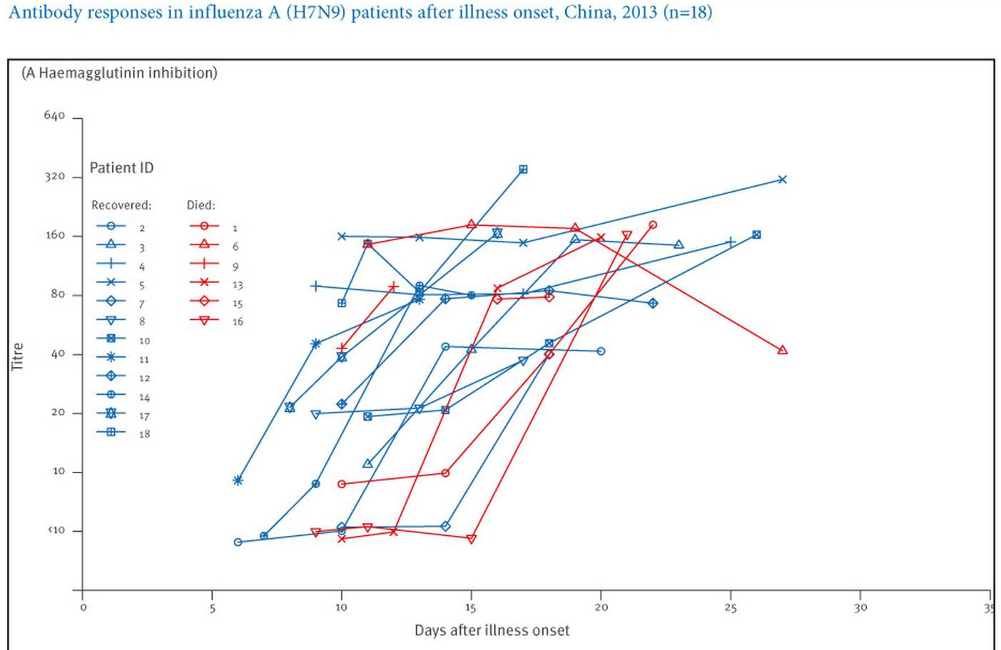
Instead, you need actual measurements using serum samples from people who are known to have been infected with H7N9 to tell you what your titers mean. Someone has to study a number of patients to see what their actual H7N9 antibody titers are, and then a mathematical analysis of that data is done to come up with a threshold titer value, above which serum samples can be said to have come from people infected with H7N9 with some large degree of certainty, and below which they are thought to have come from people who were not infected. We've seen almost no asymptomatic cases (cases with no symptoms), so we really can't say much about them. So we have to go with data from H7N9 patients who have had symptoms. Here's a great graph showing antibody titers, as measured using the HI assay, in serum samples from H7N9 patients:5

Figure 1. H7N9 HI
Euro Surveill. 2013 Dec 12;18(50):20657

As you can see in the graph above (Figure 1), by around 3 weeks after infection onset, all samples from patients whose HI titer was measured had titers 40.

The graph below (Figure 2), from a different study,4 shows that the HI titer for all H7N9 samples studied by this set of authors was also 40. In addition, this graph shows titers from "control" samples (i.e., samples from people who did not have H7N9 infections); all control samples had titers that were <40.

Figure 2: H7N9 IC50 HI4
Emerg Infect Dis. 2013 Oct;19(10):1685-7

Finally, below (Figure 3) is another nice graph, from a third study,6 showing anti-H7N9 antibody levels ("IgG"), "HI" assay results and MN assay ("NAb") results for several H7N9 patients, again showing that all samples from the H7N9 patients studied had HI titers 40. This graph also shows that all H7N9 patient serum samples had an MN titer of 20, if samples were taken after enough time had elapsed since their infections had started.

Figure 3. H7N9 IgG HI NAb.
Emerg Infect Dis. 2014 Feb;20(2):192-200

In other words, if an individual's anti-H7N9 antibody titer is 40 by the HI assay or 20 by the MN assay, these data suggest that we could pretty safely say that he or she has had a symptomatic H7N9 infection within the past few months, and if the HI or MN titers are below those cutoffs, then the individual probably hasn't had a symptomatic H7N9 infection. We don't know to what extent asymptomatic H7N9 infections will be captured by these cutoffs, but it is likely that some asymptomatic cases would be missed using these cutoffs. It is also possible that some mild infections could be missed using these cutoffs. However, it would be a great step forward just to get estimates of what percentages of any regional population or occupational group of people have had any kind of H7N9 infection. A comparison of antibody titers for asymptomatically infected and symptomatically infected H5N1 cases may be instructive when thinking about H7N9.7

WHO guidelines are even stricter than the cutoffs discussed in the paragraph above. WHO guidelines say that, using the HI assay, only single samples with titers of 160 can be considered "seropositive": "Paired sera (acute and convalescent sera) with a 4-fold rise in HI titer or single sera collected in convalescent phase with HI titer of ≥160 could be considered as H7N9 HI antibody positive. Sera with HI titer of 20-80 should be confirmed by MN or WB assay."1 For the MN assay, however, the WHO does not give specific cutoffs: "With single-serum samples, care must be taken in interpreting low titers such as 20 and 40. Generally, knowledge of the antibody titers in an age-matched control population is needed to determine the minimum titer that is indicative of a specific antibody response to the virus used in the assay."2

Now, it should be noted that WHO assay instructions recommend the use of horse red blood cells for the HI assay, and not everyone uses horse red blood cells. Some people use chicken, turkey, guinea pig or other kinds of red blood cells. That starts making comparisons between different groups' assays difficult. Horse red blood cells are better to use than turkey red blood cells for H7N9 because they have more a2,3-linked ("bird") sialic acids (influenza receptors); HI results are more sensitive with horse red blood cells. In other words, it may take less antibody in the assays to get the same result using horse red blood cells than it would using turkey red blood cells. This would translate into a higher number, when discussing H7N9 patient titers, for HI assays using horse red blood cells, compared to assays using turkey red blood cells. I have not seen direct comparisons of titers obtained using different types of red blood cells in HI assays specifically for H7N9, but the situation is probably similar to that for H5N1.8

Figures 1 and 3 above were made with HI data obtained using horse red blood cells. Figure 2 used guinea pig red blood cells. Are they completely comparable? No. Are they pretty comparable? Yes.

Are you getting a feeling for how complicated it is to interpret a seroprevalence paper? And for how difficult it is to compare results across studies?

Why does all of this matter?

It matters because some seroprevalence studies don't use appropriate cutoffs. And because it can be hard to determine even what an appropriate cutoff is when red blood cells from different species are used in an HI assay. This is where the reader has to be really careful. Cutoffs for seropositivity have been a big issue9 with H5N1 seroprevalence studies; some researchers have used cutoffs that were too low, and hence they have almost certainly overestimated how common H5N1-specific antibodies were in the populations studied.

So far, only one H7N9 serology paper published to date has reported probable seropositive samples, and this paper simply reported HI titers without using any specific threshold for seropositivity. Only one used study horse red blood cells in HI assays. The one paper that used an MN assay did use appropriate cutoffs. It should be noted that the new WHO HI guidelines were only published in December, 2013, after a couple of these papers were already published.

Here are the studies that have been published so far (I hope I haven't left any out):

  1. Bai et al.10 looked at serum samples collected before November, 2012 from poultry workers in eastern China and found no H7N9-positive samples. The study used HI and MN assays. Turkey red blood cells were used in the HI assay. Appropriate cutoffs were used for the MN assay.
  2. Hsieh et al.11 studied 14 close contacts of the first H7N9 case in Taiwan. The authors took blood samples within 18-28 days after the contacts' earliest exposures. The authors used an HI assay but not an MN assay. They used turkey red blood cells for the HI assay. They found all contacts to have an HI titer £10, and declared all to be seronegative. The HI titer for the H7N9 patient in their study was 1:80. These conclusions seem very sound.
  3. Yang et al.12 looked at serum samples from 1129 people from regions of China in which H7N9 cases had been seen, and from 396 poultry workers from 10 districts in which H7N9 cases had been seen. None of the samples from the general population was found to be seropositive, whereas >6% of the poultry workers were found to be seropositive. The authors also examined serum samples from several H7N9 patients. The study used an HI assay but not an MN assay. The authors used a cutoff of 80, along with turkey red blood cells, for the HI assay. Because the authors examined serum samples from H7N9 patients using their methods and got results that are reasonably similar to other results, their cutoffs are most likely reasonable, and their conclusions are probably quite sound. The authors report:
    • "Of the 1129 serum samples collected from individuals (age range, 1–88 years) in the general population, 9 (0.8%) had an HI titer of≥40 to influenza A(H7N9), but no serum samples with an HI titer of≥80 were found (Table 1). In contrast, among poultry workers, 13.9% (55/396) and 6.3% (25/396) had influenza A(H7N9) antibody titers of ≥40 and ≥80 (20 had an HI titer of 80, and 5 had an HI titer of 160), respectively."

      It is hard to imagine that an HI titer of 160 can be a spurious finding ("non-specific," to the initiated). Thus, these data strongly suggest that at least some H7N9 cases have been going undetected among poultry workers. Suppose we consider only the poultry workers with HI titer ≥80, or 6.3% of the poultry workers. If we then consider how many poultry workers there are, total, in districts from which H7N9 cases have emerged, then this study suggests that it's possible that quite a large number of poultry workers have been exposed to H7N9. Still, this study examined only a very small number of people, and we should be cautious about reading too much into these results.
  4. Qiu et al.13 looked at 3 H7N9 patients and 3 close household contacts of the patients who were exposed before infection control practices were put in place. The authors looked for viral RNA using a sensitive test (PCR) and examined serum samples drawn 15-26 days post-exposure using both an HI assay and a pseudovirus-based neutralization assay. They found no contacts to be seropositive. The H7N9 patients had HI titers that reached 160-640 during this time, and the patient contacts all had titers <10. The authors used horse red blood cells for the HI assay. These findings also seem sound.
To summarize, the conclusions from all of these papers do seem sound. But, it would be wise to keep all of these issues in mind as subsequent studies appear over time.

An additional study14looked at antibody titers in 1723 serum samples collected in Vietnam using a very different kind of assay (a protein microarray). Because seropositivity cutoff levels had not been determined with authors' assay methods using actual H7N9 patient samples, these authors were appropriately very careful not to attempt to draw any conclusions about H7N9 seroprevalence from their data:

"Because titers calculated from our assay are not directly comparable to HI or microneutralization tests, no cutoff is chosen to represent positivity or clinical protection. It is not possible to associate these titers with past exposure or past infection, as serological assays have not yet been validated for H7N9."

For the future

So, as new H7N9 serology studies gradually come out, you be the judge. Figure out whether they're believable or not. Ask yourself the following:
  1. What assay(s) were used? Did the authors use an MN assay? They get bonus points if they did. 
    • If only an HI assay was used, then the conclusions are slightly less certain than if an MN assay was used.
  2. If the authors used an HI assay, what species were the red blood cells from?
    • If horse red blood cells weren't used, then HI titer cutoffs lower than 160 are probably appropriate, but there is also more uncertainty about what an appropriate cutoff would be.
  3. What cutoff(s) did they use for seropositivity in their assay(s)? Do these cutoffs mesh with WHO guidelines? Do they mesh with what we know about H7N9 patient HI and MN antibody titers?
References
  1. http://www.who.int/influenza/gisrs_laboratory/cnic_serological_diagnosis_hai_a_h7n9_20131220.pdf
  2. http://www.who.int/influenza/gisrs_laboratory/cnic_serological_diagnosis_microneutralization_a_h7n9.pdf
  3. http://www.virology.ws/2009/05/27/influenza-hemagglutination-inhibition-assay/
  4. Qiu C, Huang Y, Zhang A, Tian D, Wan Y, Zhang X, Zhang W, Zhang Z, Yuan Z, Hu Y, Zhang X, Xu J. Safe pseudovirus-based assay for neutralization antibodies against influenza A(H7N9) virus. Emerg Infect Dis. 2013 Oct;19(10):1685-7
  5. Zhang A, Huang Y, Tian D, Lau EH, Wan Y, Liu X, Dong Y, Song Z, Zhang X, Zhang J, Bao M, Zhou M, Yuan S, Sun J, Zhu Z, Hu Y, Chen L, Leung CY, Wu JT, Zhang Z, Zhang X, Peiris JS, Xu J. Kinetics of serological responses in influenza A(H7N9)-infected patients correlate with clinical outcome in China, 2013. Euro Surveill. 2013 Dec 12;18(50):20657 
  6. Guo L, Zhang X, Ren L, Yu X, Chen L, Zhou H, Gao X, Teng Z, Li J, Hu J, Wu C, Xiao X, Zhu Y, Wang Q, Pang X, Jin Q, Wu F, Wang J. Human antibody responses to avian influenza A(H7N9) virus, 2013. Emerg Infect Dis. 2014 Feb;20(2):192-200
  7. Buchy P et al., PLoS One. 2010 May 27;5(5):e10864
  8. See, e.g., Table 4 in Pawar SD et al., Virol J. 2012 Oct 30;9:251
  9. Osterholm MT and Kelley NS, MBio. 2012 Feb 24;3(2):e00045-12
  10. Bai T et al., N Engl J Med. 2013 Jun 13;368(24):2339-40
  11. Hsieh SM et al., J Infect. 2013 Nov;67(5):494-5
  12. Yang S et al., J Infect Dis. 2014 Jan 15;209(2):265-9
  13. Qiu C et al., J Clin Virol. 2014 Feb;59(2):129-31
  14. Boni MF et al., J Infect Dis. 2013 Aug 15;208(4):554-8

NOTE: I did not have a hand in writing this post and thus take no credit for it. This was entirely the work of the Guest Writer. 

Wednesday, January 29, 2014

MERS-CoV antibodies in dromedary camels from Dubai, UAE, as far back as 2005...

Alexandersen and colleagues from Canada and the United Arab Emirates (UAE), writing in Transboundary and Emerging Diseases, recently described detecting antibodies to the MERS-CoV, or a close relative 

Their study is distinguished from similarly themed reports because it uses camel serum samples which are not as diluted. The thinking is that these may yield better indications of weaker positives

It also differs in that it has not undertaken all the various validation steps used in many of the antibody studies I've listed below, to convince the dubious reader that positive results are not due to some other coronavirus that may be yielding a cross-reactive and thus falsely negative result.

MERS-CoV RT-PCRs were negative on extracts of serum aliquots.

The authors could not determine where or how the camels may have been exposed to infection, other than it had been prior to the beginning of sampling in 2005. 6 camels from North America ("likely" originating from Australia) were antibody-negative reinforcing the fairly localised nature of MERS-CoV's (or it's close relative) likely origin.

So far we've read of antibodies in camels that are not convincingly present in other animals including sheep, goats, chickens, cattle, horses or camels from outside Europe, America of Australia. These antibodies react with, and sometimes neutralise the infectivity of, MERS-CoV (or a very close relative). This list now includes dromedary camels from:


With this much data behind us, camels currently sit at the top of the MERS-CoV (or some other novel CoV)-positive "animals-tested-to date" list.

Thursday, January 23, 2014

Pre-existing antibody reactive to avian influenza A(H7N9) virus did not predict better survival

Freeman and Cowling comment in the Journal of Infectious Diseases on a paper last year by Yang and colleagues (I made a note about that one here). They also re-analysed one of the conclusion and found that, for this dataset at least, having H7N9 antibodies did not afford a reduced risk of death. 

Freeman and Cowling conclude that this doesn't negate using convalescent sera (the bit of blood, minus the cells and the clotting factors, that contain proteins, water and the antibodies we make against an infection we've had) as a treatment option. But from the data in Yang's paper, the pre-infection existence of higher levels of antibodies that react with H7N9, did not improve chances for survival. 

More study is needed.

Thursday, January 2, 2014

Antibodies in 10-year old UAE camel sera suggestive, but not evidentiary, of the presence of MERS-CoV a decade ago

Click on image to enlarge. 
I've cobbled together a graphic of the assays
that have come from Prof Christian Drosten group and
colleagues, mostly for the detection and
confirmation of MERS-CoV in human samples.
632 of 651 (97.1%) dromedary camel serum samples collected in 2003 and 2013 in the United Arab Emirates (UAE) have been found to react with Middle East respiratory syndrome coronavirus (MERS-CoV) or key pieces thereof.

Meyer and colleagues from the Netherlands, Germany and the UAE also tested 16 control samples from German zoo camels but none reacted to MERS-CoV in their testing system. This indicates that the camels have not been infected by the MERS-CoV (or something very much like it) leading the authors to suggest that the virus is relatively isolated to Arabian peninsula's eastern edge...as far as we know from the testing performed to date. 


This is a potentially huge piece of good news because it suggests, to me at least, that there is a very strong chance that the spread of MERS-CoV can be contained. It will however, take a collaborative effort to "stamp out" MERS-CoV the same way SARS-CoV was stopped in its tracks (to partly quote Mike Coston) through effective infection prevention and control measures being created, implemented and enforced. 


In the absence of further testing from other regions around the world, we hold information in our hands that suggests a region-specific isolation to the MERS-CoV. And we know that right now it does not seem to be very good at all at transmitting from human-to-human. Perhaps reflecting that it is currently a camel virus and not a human one? Of course, it may never evolve into a human virus.


From what we do know today of the MERS-CoV, stamping out human infections may involve some of the following steps:


  • Being aware of the risk of contact between humans and camels and seeking to limit such contact if it could occur in the absence of suitable precautions including personal protective equipment
  • Testing camels for active infection, which may not result in notable disease in camels, and isolating those camels from other camel herds to try and "burn out" infection in camels altogether. The horse racing industry might have some good advice in this department
  • Learning more about all aspects of MERS-CoV acquisition, spread and disease in camels and perhaps in other animals. This will be influenced by future screening projects results which will hopefully identify any other animals that also a close relative/immediate ancestor that is passed to camels and then humans, or perhaps directly to humans
  •  Implementing ways to break the chain of spread from a putative other animal host to camels to people. 

Hopefully such steps could be achieved without any long term impact to camel interaction in the region as they are an essential source of social, economic and dietary enrichment.


A recombinant MERS-CoV Spike immunofluorescent assay was used to screen samples for reactive antibodies. Vero cell s expressing a recombinant Spike protein from MERS-CoV or HCoV-OC43 (used to detect cross-reactive antibodies) were fixed and then incubated with diluted animal or control serum samples (1:20 - 1:80 at 37'C for 60-min). Captured antibody was labelled with an anti-llama antibody fragment labelled with a fluorescent tag (FITC). A MERS-CoV human protein microarray assay was used to confirm screening results (I've noted this assay previously here). Virus neutralization studies were also conducted using a method I've previously written about


Meyer's study also screened 182 camel's faecal samples collected in 2013 using broad-ranging CoV RT-PCRs which, upon nucleotide sequence confirmation, yielded 2 bovine coronavirus (BCoV) positives, but no MERS-CoV positives. We learn from this that recently stored faecal samples can yield CoV RNA that can also be sequenced.


It's also worth a quick hop back to looking at the bigger picture of animal testing for a moment. Succeeding in detecting MERS-CoV RNA among the relatively small numbers of samples tested to date is akin to finding the Arkenstone among Erebor's piles of gold (even if it looked easy in the movie). Sure, a decent number of different animal species have been tested so far, but only small numbers from each. And even though there is a high proportion of camels with MERS-CoV (or its antigenic kin) antibodies, we still have to strike it lucky enough to sample during what may well be an acute virus replication period lasting only days to a couple of weeks. So far, it looks like luck has been as slippery as a woodland elf on a riverbank. Larger numbers of each animal species, camels especially, should land a hit or two in the near future I'm betting.


There is still much testing to be done, but perhaps it's possible to shut the gate before the camels have truly bolted.


Hat-tip to Helen Branswell on Twitter and her article here.

Sunday, October 27, 2013

MERS-CoV antibodies not found in children in 2010-11 or adults from 2012

Gierer and colleagues from the German primate center and the University of Dammam in Saudi Arabia, have presented the findings of their study of antibodies to the Middle East respiratory syndrome coronavirus (MERS-CoV).

The publication, in Emerging Infectious Diseases (ahead of print - you can find it here, at least until it's other link here starts working), measured the antibodies capable of blocking infection by MERS-CoV, called "neutralising antibodies" with a method they have described before. The assay was not validated with multiple MERS-CoV-positive patient sera, but appeared specific in the testing completed. None of the sera stopped the MERS-CoV Spike protein coated virus-like particles (VLP) from entering the Caco-2 cell line. Entry of the lentivirus/Spike hybrids was measured by enzyme activity inside the cells if infection is successful. Less or no activity if the VLP could not enter the cell because the VLP's Spike proteins were bound after pre-incubation with anti-MERS-CoV-containing patient sample. 

Patient samples from the area served by King Fahd Hospital were obtained from:

  1. Children (158 sera, 77 female, mean age 12 months) admitted to hospital with lower respiratory tract infections during 12-months form May 2010. 
  2. Adult (110 plasma samples, all males, mean age 28-years, upper limit of 52-years) blood donors 
No sera or plasma had neutralising MERS-CoV antibodies.

The authors conclude that <2.3% of children and <3.3% pod adults were seropositive though, because that accounts for the upper limit of the confidence intervals. They also note that their sampling of hospitalized children could have missed an antibody response (because it takes time to develop) if they had only just been admitted to hospital for MERS-CoV. 

Additionally, its a pretty small sample on which to be base too many conclusions when considering a virus that is spread across a 2,100,000kms2 and reportedly caused notable disease in <200 of 28,000,000 people.


Thursday, September 5, 2013

More MERS molecular masterfulness: Egyptian camels contain lots of anti-MERS-CoV antibodies [AMENDED]

Perera and colleagues from China, Japan, Egypt and the United States report in Eurosurveillance that they
have found a high prevalence of Egyptian camels with antibodies to a piece of the Middle East respiratory syndrome coronavirus (MERS-CoV) spike protein.

The group looked at 1,343 human sera (815 from Cairo as part of influenza study; 528 archived from Hong Kong) and 625 animal sera (from goats, sheep, water buffalo, cows, camels, pigs and birds) from Egypt and China (no MERS reported there, so this acts as control population; unfortunately no camels tested from there either). A positive control serum was provided by Prof Drosten from an earlier MERS case.

The novel assay does not need to be used in a biosafety level 3 containment environment (BSL2 is fine) because it does not use whole/live MERS-CoV to capture the human/animal antibodies, rather it uses a piece of the virus; the Spike protein, which is known to attract the lion's share of antibody attention. The Spike protein was merged with HIV (non-infectious) proteins to create a pseudoparticle of proteins that could enter cells, and also bind to MERS-CoV antibody which would prevent entry by the virus-like particle. If there was no antibody, the pseudoparticles could enter cells and this entry could be measured using a marker enzyme reaction. This novel assay (pseudoparticle neutralization test; ppNT) was run in parallel with a more standard micro-neutralization test (MNT; the two tests agreed well but the standard NT needed to be carried out in a BSL3 laboratory), The more traditional MNT pre-incubated infectious MERS-CoV with serum and then measured whether the live MERS-CoV could infect and damage cells. If there was antibody in the serum, it blocked infection (as it can in us). Pseudoparticle/virus will be more or less blocked from cell entry if more or less antibody is present in the serum. So one can determine how much antibody was in the serum sample too. These type of tests measure the ability of antibody in a patient/animal to block virus entry; it's neutralizing ability. 

A high proportion (103/110; 93.6%) of dromedary camels from Egypt had antibodies that could block cell culture infection by a lab strain of the MERS-CoV (MERS-CoV/EMC form Erasmus University Medical Center). 

No humans had antibodies. No animal sera from China (Hong Kong) were positive - this included pigs (n=260) and wild birds (n=204). 

Numbers were sometimes very low but no goats (n=13), sheep (n=5), water buffalo (n=8) or cows (n=25) were antibody-positive in Egypt. Only the camels.

The authors specify that this does not exclude cross-reactivity with a closely related, but non-MERS, CoV that has at some time infected the camels. If only there was some sort of plan to do this sort of virus hunting in mammals.
Click to enlarge.

So how does this fit in with the human acquisition model proposed 1-week ago? Pretty well. It supports previous findings from Omani camels too. Camels could be part of the chain of infection. It says nothing about how they may then go on to be a source of human infections that start of as a respiratory illness, presumably requiring, in most cases, inhalation or self-inoculation (I'm looking at you, nose-pickers) to begin with. For hat we'd need to know anything about survival do MERS-CoV in excreta and on surfaces. I'd also like to see some more animal test results. 

Nonetheless, it looks as though we have some very useful antibody detection assays shaping up to do just that.

Here's hoping for a KSA study next.

Thursday, August 15, 2013

H5N1 did not transmit easily between humans in the wild...

Hat tip to @Laurie_Garrett and CIDRAP

Despite wearing next to no personal protective equipment (5% of 419 contacts used a mask, face shield, gown or gloves) and coming into contact with sick or dead poultry (12% of contacts), 85/87 household members and 332 "less close" contacts of 23 influenza A(H5N1) virus cases did not show any significant sign of antibodies to the virus, a study published in PLOS|ONE by Bai and colleagues noted.

Only 2 (0.4% of all contacts tested) were defined as infected by H5N1  during the study period of 2005-2008, on mainland China. 

The study used both haemagglutination test (antibodies in the patients sera bind horse red blood cells together giving a distinctive pattern) and micro-neutralization (presence of specific antibodies in a sample prevents a lab stock of virus from infecting a cell line-amount of virus can be determine by making dilutions of  the sample and comparing to a sample with no antibodies to the virus). When they had a single serum, the authors used:

  • A neutralizing antibody cut-off titre of ≥40 children (<14-years of age)  with a haemagglutination titre ≥40.
  • A neutralizing antibody cut-off titre ≥80 for those aged 15-59-years with a haemagglutination titre ≥40.
For acute and convalescent sera pairs positivity to H5N1 was defined as:

  • 4-fold rise in neutralizing antibody titre between acute and convalescent sera
  • Convalescent sera needed a neutralizing titre of ≥40 for children and ≥80 for adults, or a haemagglutination titre ≥40
There were a few more positives below these cut-offs.

While genetically altered H5N1 can be made to spread among ferrets in the lab, it seems that some years ago in the wild, H5N1 had a ways to go before it could spread efficiently between humans. That's a good thing.